Radiology is a medical specialty that uses imaging techniques to diagnose and treat diseases within the body. It encompasses various modalities, including X-rays, computed tomography (CT) scans, magnetic resonance imaging (MRI), ultrasound, and nuclear medicine.
Key Imaging Techniques:
X-rays: Produce images of bones and dense tissues by passing radiation through the body.
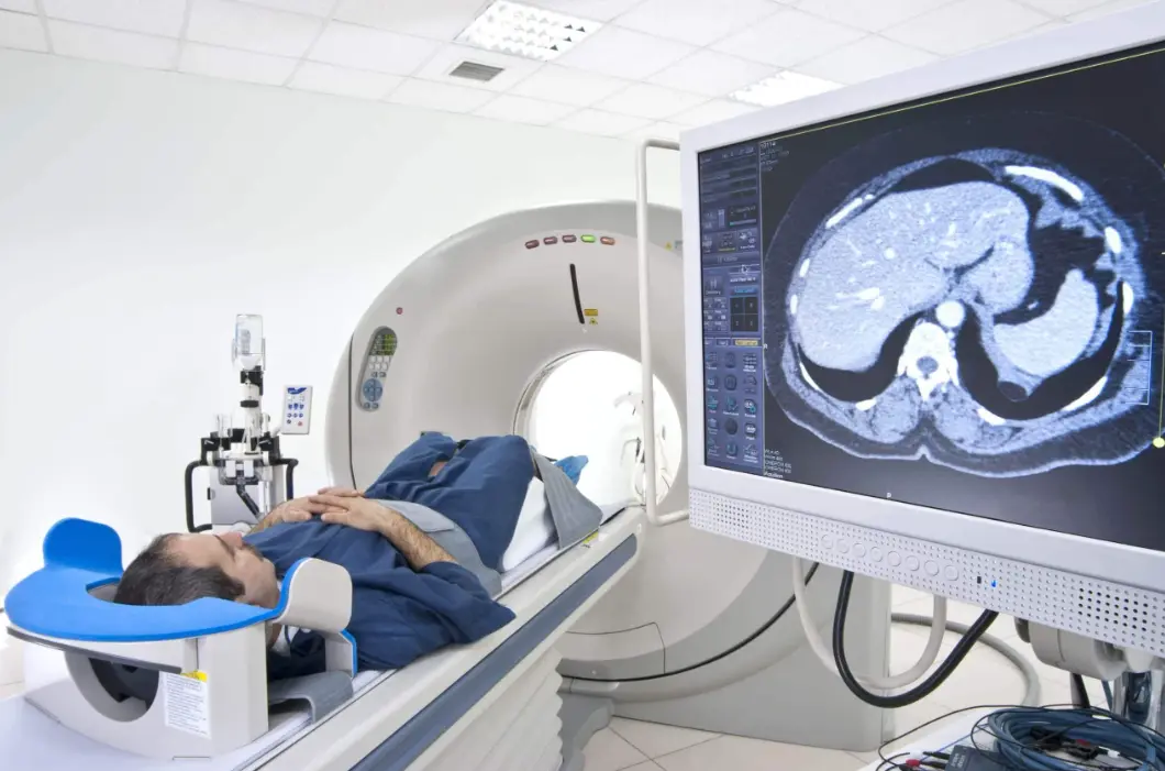
CT Scans: Provide detailed cross-sectional images using X-rays and computer processing.
MRI: Uses magnetic fields and radio waves to create images of soft tissues, organs, and the brain.
Ultrasound: Employs sound waves to visualize internal structures, commonly used in obstetrics and cardiology.
Nuclear Medicine: Involves radioactive substances to assess organ function and detect abnormalities.
Applications in Medicine:
Radiology plays a crucial role in early detection of conditions such as fractures, tumors, infections, and cardiovascular diseases. It aids in guiding minimally invasive procedures, monitoring treatment progress, and planning surgeries. Subspecialties include interventional radiology, which uses imaging for targeted therapies like stent placements or tumor ablations.
Importance and Advances:
Advancements in technology, such as artificial intelligence and 3D imaging, have enhanced diagnostic accuracy and reduced radiation exposure. Radiology teams, including radiologists and technicians, collaborate with other healthcare professionals to improve patient outcomes and personalize care.
Table of contents
- Part 1: OnlineExamMaker – Generate and share radiology quiz with AI automatically
- Part 2: 20 radiology quiz questions & answers
- Part 3: Automatically generate quiz questions using AI Question Generator
Part 1: OnlineExamMaker – Generate and share radiology quiz with AI automatically
The quickest way to assess the radiology knowledge of candidates is using an AI assessment platform like OnlineExamMaker. With OnlineExamMaker AI Question Generator, you are able to input content—like text, documents, or topics—and then automatically generate questions in various formats (multiple-choice, true/false, short answer). Its AI Exam Grader can automatically grade the exam and generate insightful reports after your candidate submit the assessment.
What you will like:
● Create a question pool through the question bank and specify how many questions you want to be randomly selected among these questions.
● Allow the quiz taker to answer by uploading video or a Word document, adding an image, and recording an audio file.
● Display the feedback for correct or incorrect answers instantly after a question is answered.
● Create a lead generation form to collect an exam taker’s information, such as email, mobile phone, work title, company profile and so on.
Automatically generate questions using AI
Part 2: 20 radiology quiz questions & answers
or
1. What is the primary advantage of using MRI over CT scans for brain imaging?
A. Faster imaging time
B. Better visualization of soft tissues without ionizing radiation
C. Lower cost
D. Ability to image bones more clearly
Answer: B
Explanation: MRI uses magnetic fields and radio waves, providing superior soft tissue contrast without the risks associated with ionizing radiation from CT scans.
2. In a chest X-ray, what does hilar lymphadenopathy typically indicate?
A. Lung infection
B. Heart enlargement
C. Possible malignancy or infection in the lymph nodes
D. Rib fractures
Answer: C
Explanation: Hilar lymphadenopathy appears as enlarged lymph nodes in the hilar region, often suggesting conditions like tuberculosis, sarcoidosis, or lung cancer.
3. Which imaging modality is most commonly used to detect gallstones?
A. MRI
B. Ultrasound
C. CT scan
D. X-ray
Answer: B
Explanation: Ultrasound is preferred for gallstone detection due to its non-invasive nature, lack of radiation, and ability to visualize the gallbladder in real-time.
4. What is the main purpose of a barium swallow study?
A. To evaluate the stomach
B. To assess the esophagus for structural abnormalities
C. To image the small intestine
D. To detect kidney stones
Answer: B
Explanation: A barium swallow involves ingesting barium sulfate to coat the esophagus, allowing X-ray visualization of swallowing disorders, strictures, or tumors.
5. In mammography, what does a microcalcification cluster suggest?
A. Benign fibroadenoma
B. Possible early breast cancer
C. Cystic changes
D. Fatty tissue accumulation
Answer: B
Explanation: Microcalcifications on mammograms can indicate ductal carcinoma in situ or invasive breast cancer, prompting further biopsy for confirmation.
6. Which contrast agent is commonly used in CT scans for vascular imaging?
A. Gadolinium
B. Iodine-based contrast
C. Barium sulfate
D. Technetium-99m
Answer: B
Explanation: Iodine-based contrast enhances blood vessels on CT scans by increasing attenuation, making it ideal for angiography and detecting vascular abnormalities.
7. What artifact might appear on an MRI due to patient movement?
A. Ghosting
B. Ring artifact
C. Beam hardening
D. Partial volume effect
Answer: A
Explanation: Patient movement during MRI acquisition can cause ghosting artifacts, which appear as blurred or duplicated images, degrading diagnostic quality.
8. In pediatric radiology, why is ultrasound often preferred over CT?
A. It provides better bone detail
B. It avoids ionizing radiation
C. It is faster
D. It requires sedation less often
Answer: B
Explanation: Ultrasound uses sound waves and does not involve radiation, making it safer for children to reduce long-term cancer risk from repeated imaging.
9. What is the typical finding on a CT scan for a pulmonary embolism?
A. Ground-glass opacity
B. Filling defect in the pulmonary artery
C. Pleural effusion
D. Hyperinflated lungs
Answer: B
Explanation: CT pulmonary angiography shows a filling defect, indicating a blood clot obstructing the pulmonary artery, which is diagnostic for pulmonary embolism.
10. Which X-ray view is best for evaluating the knee joint?
A. Anteroposterior (AP) view
B. Lateral view
C. Oblique view
D. Both A and B
Answer: D
Explanation: Both AP and lateral views are essential for assessing knee alignment, fractures, and joint space, providing a comprehensive evaluation.
11. In nuclear medicine, what does a bone scan primarily detect?
A. Soft tissue infections
B. Bone metastases or fractures
C. Brain tumors
D. Liver function
Answer: B
Explanation: Bone scans use radiotracers to highlight areas of increased bone activity, such as metastases, infections, or fractures, by detecting abnormal uptake.
12. What is the risk associated with repeated CT scans?
A. Increased radiation exposure leading to cancer
B. Allergic reactions to contrast
C. Motion artifacts
D. Poor image resolution
Answer: A
Explanation: CT scans involve ionizing radiation, and cumulative exposure can elevate the risk of developing radiation-induced cancers over time.
13. Which imaging technique is used for real-time guidance in biopsies?
A. Plain X-ray
B. Fluoroscopy
C. MRI
D. Ultrasound
Answer: D
Explanation: Ultrasound provides dynamic, real-time imaging without radiation, making it ideal for guiding needle biopsies of organs like the liver or thyroid.
14. On an abdominal X-ray, what does a kidney-ureter-bladder (KUB) view show?
A. Gallbladder stones
B. Urinary tract stones or obstructions
C. Liver lesions
D. Pancreatic inflammation
Answer: B
Explanation: A KUB X-ray visualizes the kidneys, ureters, and bladder, commonly used to detect renal calculi or bowel gas patterns indicating obstruction.
15. What does diffusion-weighted imaging (DWI) on MRI indicate in stroke patients?
A. Hemorrhagic stroke
B. Ischemic stroke
C. Tumor growth
D. Vascular malformations
Answer: B
Explanation: DWI on MRI shows restricted diffusion in ischemic areas due to cytotoxic edema, allowing early detection of acute strokes within hours.
16. In interventional radiology, what is the purpose of angioplasty?
A. To remove tumors
B. To open blocked blood vessels
C. To biopsy tissues
D. To drain abscesses
Answer: B
Explanation: Angioplasty uses a balloon catheter to widen narrowed or obstructed arteries, improving blood flow and treating conditions like atherosclerosis.
17. Which modality is least suitable for imaging patients with pacemakers?
A. Ultrasound
B. MRI
C. CT scan
D. X-ray
Answer: B
Explanation: MRI’s strong magnetic fields can interfere with pacemakers, potentially causing device malfunction, making it contraindicated in most cases.
18. What is a common indication for a PET-CT scan?
A. Detecting bone density
B. Staging cancer and assessing metastases
C. Evaluating heart function
D. Imaging the eyes
Answer: B
Explanation: PET-CT combines metabolic and anatomical imaging to detect cancer spread, monitor treatment response, and identify metabolically active tumors.
19. In a head CT scan, what might hyperdense areas in the brain represent?
A. Edema
B. Hemorrhage
C. Infarction
D. Atrophy
Answer: B
Explanation: Hyperdense areas on CT indicate acute blood, as in hemorrhage, due to the high attenuation of blood compared to surrounding brain tissue.
20. Which factor increases the risk of contrast-induced nephropathy?
A. Young age
B. Pre-existing kidney disease
C. High blood pressure
D. Mild dehydration
Answer: B
Explanation: Patients with pre-existing renal impairment are at higher risk for contrast-induced nephropathy when using iodinated contrast agents in procedures like CT.
or
Part 3: Automatically generate quiz questions using OnlineExamMaker AI Question Generator
Automatically generate questions using AI